Medlea
LeaRes · functional digital twin

The patient’s anatomy,
as a working model.

LeaRes turns a chest CT into a patient-specific functional twin of the lungs: anatomy reconstructed, ventilation and airflow simulated, in the browser, on your hospital’s GPU.

  • Lung anatomy
  • AI segmentation
  • Ventilation simulation
  • Virtual spirometry (RUO)
Virtual spirometry · forced expirationDemo case
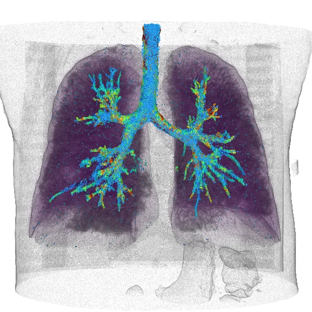
Both lungs reconstructed from a chest CT during a simulated forced expiration
FEV1
3.20 L
FVC
4.38 L
FEV1/FVC
72.9 %

All MedLea solutions are browser-accessible and comply with GDPR · HIPAA · SOC2 · EU AI Act.

  • GDPR
  • HIPAA
  • SOC2
  • EU AI Act

LeaRes is CE-marked medical device software (class I) for study, simulation and the training of healthcare professionals. It does not provide a diagnosis: the judgement stays with the clinician.

01
Morphology and airways

Anatomy that breathes

Lobes and airway tree reconstructed from the CT by AI segmentation, then set in motion: airflow simulated in every airway, regional ventilation in every lobe, on the patient’s own anatomy.

  • Five lobes, trachea and bronchial tree, segmented case by case
  • Airflow speed along the tree, from a GPU flow solver
  • Scenarios compared on one patient-specific model, for study and teaching
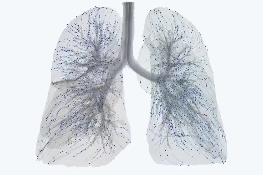
Airflow in the airways and parenchyma
Anatomy that breathes
02
Reinflation and hyperdistension

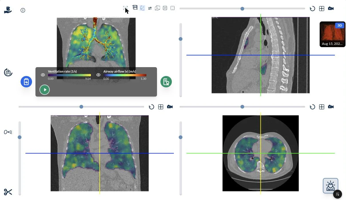
Regional ventilation, slice by slice

The breathing twin yields regional biomechanics: where the lung inflates, where it over-distends, how the two lungs share the breath. Simulated maps you can read on the familiar MPR views, for research and training.

  • Ventilation rate mapped onto axial, coronal and sagittal slices
  • Simulated FEV1 and FVC per lobe (virtual spirometry, research use only)
  • Simulation outputs for study, never a clinical verdict
Airflow and biomechanics of the airways and parenchymaDemo case
Regional ventilation, slice by slice
03
Research & development · blood flow

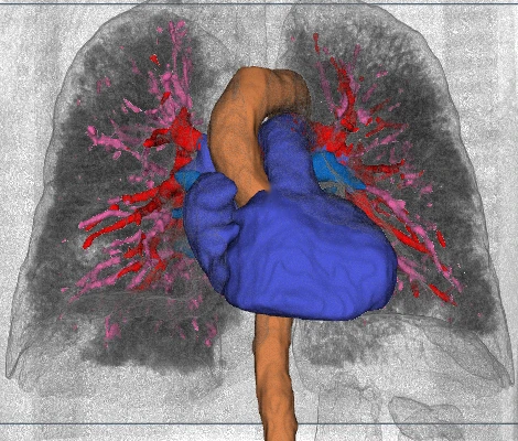
The vessels, from the same scan

In development at Medlea, not a function of the CE-marked device: pulmonary arteries and veins, heart and aorta reconstructed alongside the lungs, as the geometry a flow study needs for perfusion research.

  • Vascular tree and great vessels as separate, labelled structures
  • One coordinate frame with the airways and lobes
  • Exportable geometry for simulation and CAD
Heart, aorta, pulmonary arteries and veinsDemo case
The vessels, from the same scan

Engineering · research & development

From anatomy to CFD, in the same workstation

The engineering workspace takes the reconstructed anatomy, or an imported CAD geometry, sets materials and boundary conditions, and runs the flow. The full solver for the final answer; surrogates for the exploration.

  • 01

    Patient-specific CFD

    A lattice-Boltzmann multiphysics solver on GPU computes airflow, pressure and particle transport in the patient’s own airways and vessels.

  • 02

    CFD surrogates

    AI surrogates trained on full CFD runs return the same quantities in a fraction of the time, so settings and device variants can be explored interactively before the full solver confirms the final case.

  • 03

    Devices in real anatomy

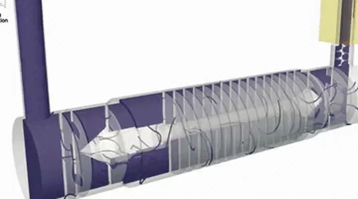
    Catheters, stents, flow diverters and ventilator circuits placed in the reconstructed anatomy or imported as CAD, with inlets, outlets and materials set in the browser.

Device CFD · streamlines in a ventilator circuit
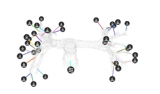
Set-up in LeaRes · materials, 35 inlets and outletsDemo case
Set-up in LeaRes · materials, 35 inlets and outlets

Active Learning · pilot programme

A model your department trains on its own cases

Name the pattern, annotate inside LeaRes, train on your GPU, correct what the model proposes. Each round leaves less to correct. The data never leaves the hospital.

How the platform works
  1. 01
    Define the pattern
  2. 02
    Annotate
  3. 03
    Train on your GPU
  4. 04
    Correct the proposal
RESEARCH & ENGINEERING · NOT PART OF THE CE-MARKED DEVICE

LeaFlow

An agentic workflow that takes a scan through segmentation, reconstruction and simulation.

LeaFlow is Medlea’s research and engineering workflow. AI agents carry a CT or MRI scan through segmentation, anatomical reconstruction and simulation set-up, step by step. At its core sits a multiphysics simulation engine: it models how blood, air, urine, drugs, contrast agents and particles move through an anatomy, and how a medical device — catheter, implant, inhaler, stent — changes that flow, so a device maker can test a prototype in a virtual anatomy before building it. LeaFlow is not intended for clinical decisions on individual patients.

Applications
04

Respiratory

Aerosol therapy, nebulisers and smart inhalers: LeaFlow injects the spray of an inhaler and follows the particles through the airways. A regional deposition model is in development.

  • Endotracheal tubes and bronchial blockers: how they change airflow and regional stress.
  • Results that support research on ventilation and the design of inhaled therapies.
Respiratory
05

Cardiovascular

Stents, grafts and valves: LeaFlow measures their effect on local flow, wall shear stress and downstream perfusion, inside the vessels of the individual patient.

  • Wall shear stress, residence times and particle transport, for device research.
  • For virtual prototyping of new devices.
Cardiovascular
06

Renal and urological

Ureteral stents, catheters and artificial sphincters: LeaFlow shows how they change urinary flow, pressure gradients and the transport of sediments and particles.

  • Encrustation, reflux and partial obstruction, studied in virtual anatomies.
  • Prototypes tested in virtual anatomies, with fewer animal models and physical builds.
Renal and urological
07

Anatomical reconstruction

CT and MRI scans become accurate 3D models: airways, vessels, renal tracts and other flow-relevant anatomy are identified and reconstructed automatically, case by case.

  • Captures both the large structures and the fine detail a simulation depends on.
  • One model serves the simulation, the CAD export and the browser viewer.
Anatomical reconstruction
08

One workflow for clinicians and engineers

Patient models move in and out of standard CAD tools. Engineers place a stent, valve or catheter in a reconstructed anatomy and test the design.

  • Export to CAD for geometric editing, structural analysis or 3D printing.
  • Imaging, simulation and design in one cycle, shared by clinical and engineering teams.
One workflow for clinicians and engineers
09

In the cloud or on premises, from the browser

LeaFlow runs in the cloud or on premises, on GPUs, for real-time or batch simulations, and every step is driven from an ordinary web browser.

  • An API connects it to research pipelines, training simulators and validation workflows.
  • Velocity fields, deposition maps, pressure gradients and residence times, in a visual interface.